

For decades, the overarching consensus in paleontology held that soft tissues, biological fluids, and delicate cellular structures were doomed to rapid decay following an organism’s death. When studying creatures that vanished tens or hundreds of millions of years ago, scientists expected to find only the hardened, heavily mineralized remnants of skeletal structures: petrified bones, teeth, or trace fossils like footprints left in ancient mud.
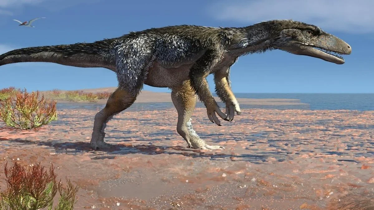
However, a landmark discovery has dramatically altered this long-held paradigm. An international research team, led by scientists at Curtin University, has uncovered fossilized remnants of red blood cells trapped deep inside the leg bone of a dinosaur. The fossil, belonging to the genus Aratasaurus, was recovered in Brazil and dates back approximately 115 million years to the Cretaceous period.
The breakthrough findings, published in the Proceedings of the National Academy of Sciences (PNAS), provide direct physical evidence that microscopic organic structures can endure far longer than previously thought possible. Beyond its immediate paleontological significance, the discovery reshapes how researchers view the fossilization process and redefines the boundaries of what information can be extracted from Earth’s deep geological record.
Cellular Micro-Traces Uncovered Deep Within Fossilized Bone
The discovery began when researchers initiated a high-resolution microscopic examination of the Aratasaurus leg bone. While analyzing the internal architecture of the fossilized osseous tissue, the team navigated through a complex, microscopic network of fine vascular canals—the tiny pathways through which blood vessels once flowed to nourish the living bone.
Deep inside these ancient vascular channels, scientists observed distinct oval, cell-like structures. The size and elliptical morphology of these shapes immediately caught the team’s attention, as they closely mirrored the red blood cells, or erythrocytes, seen in modern birds and reptiles. Unlike mammalian red blood cells, which lack cell nuclei, avian and reptilian red blood cells are typically oval and nucleated.
However, in paleontology, visual similarity alone is insufficient to claim a major biological discovery. Mineral deposits, inorganic crystal formations, or environmental micro-fossils can easily mimic the shapes of biological cells under a microscope. Recognizing the potential for false positives, the research team refrained from relying purely on structural appearances and instead initiated a comprehensive battery of micro-chemical and geochemical analyses to determine the exact origin of the microscopic shapes.
Ruling Out Contamination: Confirming a Genuine Biological Origin
Whenever potential soft tissue or cellular structures are detected in ancient fossils, the primary scientific hurdle is ruling out secondary contamination. Skeletal material buried in sedimentary rocks for over 100 million years is continually exposed to ground fluids, soil microbes, fungi, and modern environmental contamination. Fungal hyphae, bacterial biofilms, or invasive organic matter can easily penetrate porous fossil bones, creating deceptive signals during chemical testing.

To address this challenge, the international research team subjected the samples to rigorous chemical screening. The tests specifically looked for biological markers indicative of modern or historical bacterial and fungal intrusion. The chemical spectra revealed no signatures of fungal hyphae or bacterial colonization within the examined micro-canals.
Furthermore, the team analyzed the organic chemical profile of the cellular structures and compared it directly to the surrounding matrix, including the microbial covers and mineral mats that coated the outer surfaces of the bone. The chemical signature of the internal cell-like structures was distinctly different from the external microbial layer, demonstrating that the structures were intrinsic to the bone’s internal vascular system rather than introduced from the outside environment after burial.
The definitive proof came with the identification of a crucial biomarker: "heme B" molecules localized within the vascular channels. Heme B is a key iron-bearing complex that forms the core of hemoglobin, the essential protein responsible for transporting oxygen throughout the circulatory systems of animals. Finding chemical traces of heme B tightly bound within the cellular remnants inside the bone canals provided indisputable evidence. The structures were confirmed not to be mineral artifacts or microbial invaders, but the mineralized remnants of the Aratasaurus’s own red blood cells.
How Did Dinosaur Blood Cells Survive for 115 Million Years?
The survival of delicate biological structures across 115 million years of geological time requires an extraordinary alignment of geochemical and environmental factors. Under normal conditions, autolysis (cell self-destruction) and microbial decomposition dismantle soft tissues within days or weeks of an animal’s death. For red blood cells to leave a recognizable trace over millions of years, the ordinary rules of taphonomy—the process of decay and fossilization—must be interrupted almost immediately.
According to the study, the preservation of the Aratasaurus blood cells began with the rapid burial of the animal shortly after its death. The dinosaur was swiftly covered by shallow, fine-grained sediments in a low-oxygen (anoxic) microenvironment. This oxygen-depleted setting severely restricted the activity of scavengers and aerobic bacteria, significantly slowing down the initial rates of organic decomposition.
The critical phase of preservation, however, involved a specific mineral substitution process driven by a manganese oxide mineral known as hausmannite. As ground fluids rich in dissolved minerals percolated through the low-oxygen sediment, hausmannite began to precipitate directly onto the decaying red blood cells.
Rather than destroying the cells, the mineral acted as a precise microscopic casting agent. Hausmannite coated and infused the outer boundaries of the blood cells, taking on their exact shape and structural geometry at a microscopic scale. As the original organic material slowly degraded over geological epochs, the manganese mineral solidified into a durable, permanent template.

This process created an exact mineral replica—a microscopic cast—of the dinosaur’s red blood cells that successfully endured millions of years of heat, pressure, and chemical exposure within the earth. This intricate sequence explains why such discoveries are exceedingly rare: it requires rapid burial, an anoxic environment, specific mineral chemistry, and microscopic mineral precipitation occurring simultaneously before cellular decay erases the structures entirely.
Beyond Cloning: Decoding Ancient Physiology and Space Exploration
The discovery of dinosaur blood remnants inevitably sparks public curiosity regarding the possibility of extracting intact DNA to clone extinct animals—a concept popularized by science fiction. However, researchers emphasize that the primary goal of this study, and of soft-tissue paleontology as a whole, has nothing to do with cloning. DNA is a fragile molecule that degrades relatively quickly after death, and even under optimal preservation conditions, it cannot survive intact for 115 million years.
Instead, the true value of the discovery lies in its potential to unlock fundamental questions about dinosaur biology, physiology, and evolutionary history. By analyzing mineralized cell structures and biomolecular remnants like heme B, scientists can gain unprecedented insights into how ancient creatures actually functioned internally:
- Metabolic Rates: Examining the size, structure, and density of red blood cells can help paleobiologists evaluate the metabolic demands of dinosaurs, shedding light on the long-standing debate over whether specific groups were warm-blooded (endothermic), cold-blooded (ectothermic), or somewhere in between.
- Circulatory Efficiency: Understanding the micro-anatomy of dinosaur blood channels provides direct clues regarding oxygen transport capacity, cardiovascular pressure, and overall stamina.
- Evolutionary Transitions: Comparing ancient cellular structures with those of modern birds and reptiles allows researchers to trace the step-by-step evolutionary transformation of metabolic and respiratory systems over vast geological periods.
Intriguingly, the implications of this discovery extend far beyond Earth and into the field of astrobiology. The realization that delicate organic structures can leave micro-mineralized casts capable of persisting for over 100 million years provides a vital blueprint for scientists searching for past life on other planets, such as Mars.
Robotic rovers currently exploring Martian lake beds and sedimentary basins are actively looking for potential biosignatures left behind during Mars’s ancient, water-rich past. If microscopic life once existed on the Red Planet, its physical traces may not persist as soft organic matter, but rather as mineralized micro-casts preserved by manganese or iron oxides, identical to the mechanism that preserved the blood cells of Aratasaurus.
By studying how Earth’s minerals preserve microscopic biological structures under extreme conditions, paleontologists and astrobiologists are jointly developing the diagnostic tools needed to identify ancient signatures of life, whether buried in the rocks of Brazil or in the dusty sediments of Mars.